
OUR MISSION:
Engineering translational and physiologically relevant preclinical cancer models to advance women’s health research
Research
In addition to Sanford Research, we acknowledge support from the following programs:


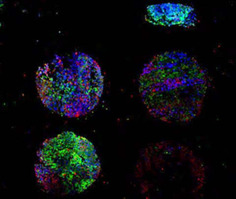
Tumor Microarray with tissue and tumoroids grown inside 3D cultures
Patient-derived tumor models as avatars for personalized prediction of therapeutic efficacy
Despite significant improvements in research and development in the cancer field, oncology clinical trials have the highest failure rate (96.6%) compared with any other therapeutic area. There is a lack of suitable preclinical models that accurately represent tumor behavior ex vivo (“out of the living”). To better understand the role and the complexity of the tumor microenvironment in human tumors, we are developing patient-derived tumors models as avatars for personalized prediction of therapeutic efficacy. This personalized drug screening platform allows rapid prediction of therapeutic response in vitro, thereby ensuring drug efficacy for each patient.
Calar K*, Plesselova S*, Bhattacharya S, Jorgensen M, De la Puente P. Human Plasma-Derived 3D Cultures Model Breast Cancer Treatment Responses and Predict Clinically Effective Drug Treatment Concentrations. Cancers (Basel). 2020 Jun 29;12(7):1722. doi: 10.3390/cancers12071722. PMID: 32610529 (* equal contribution).
Engineering immune-cancer crosstalk and physiologically relevant oxygen gradients and ECM remodeling
One of the major components of the tumor microenvironment is the extracellular matrix (ECM), which undergoes remodeling and contributes to immune evasion mechanisms. Low oxygen is a key regulator of ECM remodeling, however, little is known about the role of the hypoxia-induced ECM remodeling in immune evasion and response to immunotherapy. We use patient-derived 3D model quantitative research methods to gain insight into the recreation of the hypoxia-induced ECM remodeling and its role in immune evasion.
Bhattacharya S, Calar K, de la Puente P. Mimicking tumor hypoxia and tumor-immune interactions employing three-dimensional in vitro models. J Exp Clin Cancer Res. 2020 May 1;39(1):75. doi: 10.1186/s13046-020-01583-1. PMID: 32357910
Bhattacharya S, Calar K, Evans C, Petrasko M, de la Puente P. Bioengineering the oxygen-deprived tumor microenvironment within a three-dimensional platform for studying tumor-immune interactions. Front Bioeng Biotechnol. 2020 Sep 4;8:1040. doi: 10.3389/fbioe.2020.01040. PMID: 33015012
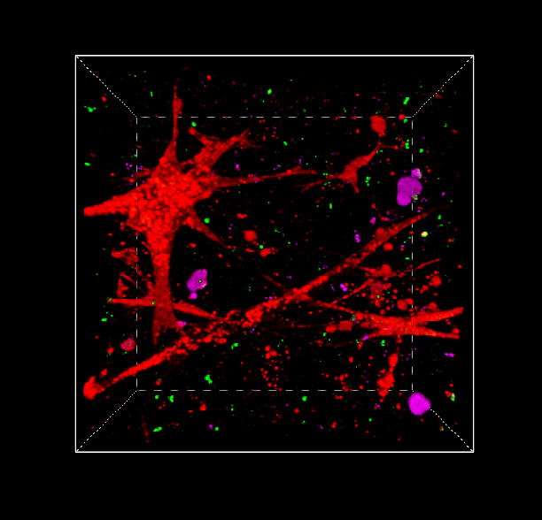
Z-stack of 3D tri-culture of cancer, cancer associated fibroblasts and immune cells
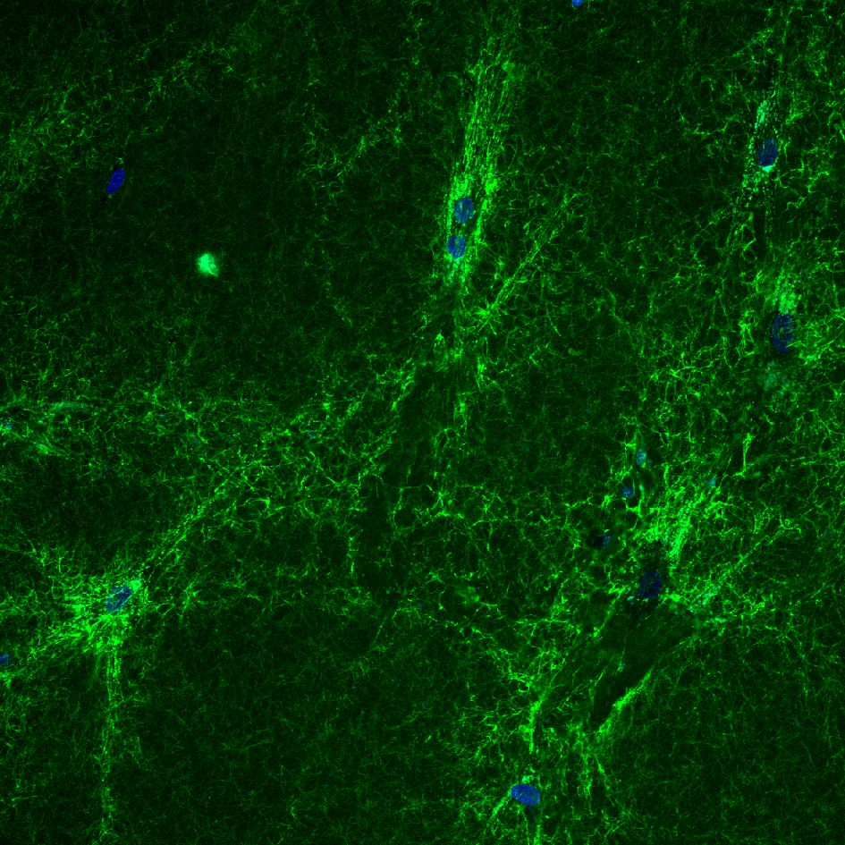
Collagen expression by cancer-associated fibroblasts in 3D culture
Reprogramming normal fibroblasts into cancer-associated fibroblast
Cancer-associated fibroblasts (CAFs) are key contributors to cancer progression and therapeutic resistance through dysregulation of the extracellular matrix (ECM). CAFs are a heterogenous population derived from different cell types through activation and reprogramming. Current studies rely on uncharacterized heterogenous primary CAFs or normal fibroblasts that fail to recapitulate CAF-like tumor behavior. Our lab has developed a translational based approach to reprogram normal fibroblasts into CAFs using conditioned media from cancer lines. Using these resources, further development of therapeutics that possess potentiality and specificity towards CAF/ECM-mediated chemoresistance in OC are further warranted.
Axemaker H, Plesselova S, Calar K, Jorgensen M, Wollman J, de la Puente P. Reprogramming of normal fibroblasts into ovarian cancer-associated fibroblasts via non-vesicular paracrine signaling induces an activated fibroblast phenotype. Biochim Biophys Acta Mol Cell Res. 2024 Jul 20;1871(7):119801. doi: 10.1016/j.bbamcr.2024.119801. PMID: 39038611
Biomarkers for early detection of Ovarian Cancer
Ovarian Cancer (OC) is a deadly disease for women, known as the “silent killer” due to its few symptoms and resistance to treatment that causes disease recurrence in more than three-quarters of patients. To date there is no approved biomarker for screening of OC or clearly identified drives of tumor recurrence. Our recent studies have identified new biological makers as potential candidate biomarkers for OC and key drivers of treatment resistance. Our goal is to develop a non-invasive blood test capable of detection of OC and further characterize the biomarkers’ implication in drug resistance. These studies have the potential to lead to biomarker tests able to detect OC early and develop treatment options for OC, capable of improving chemotherapy efficacy, preventing tumor recurrence, and thus improving patient survival.
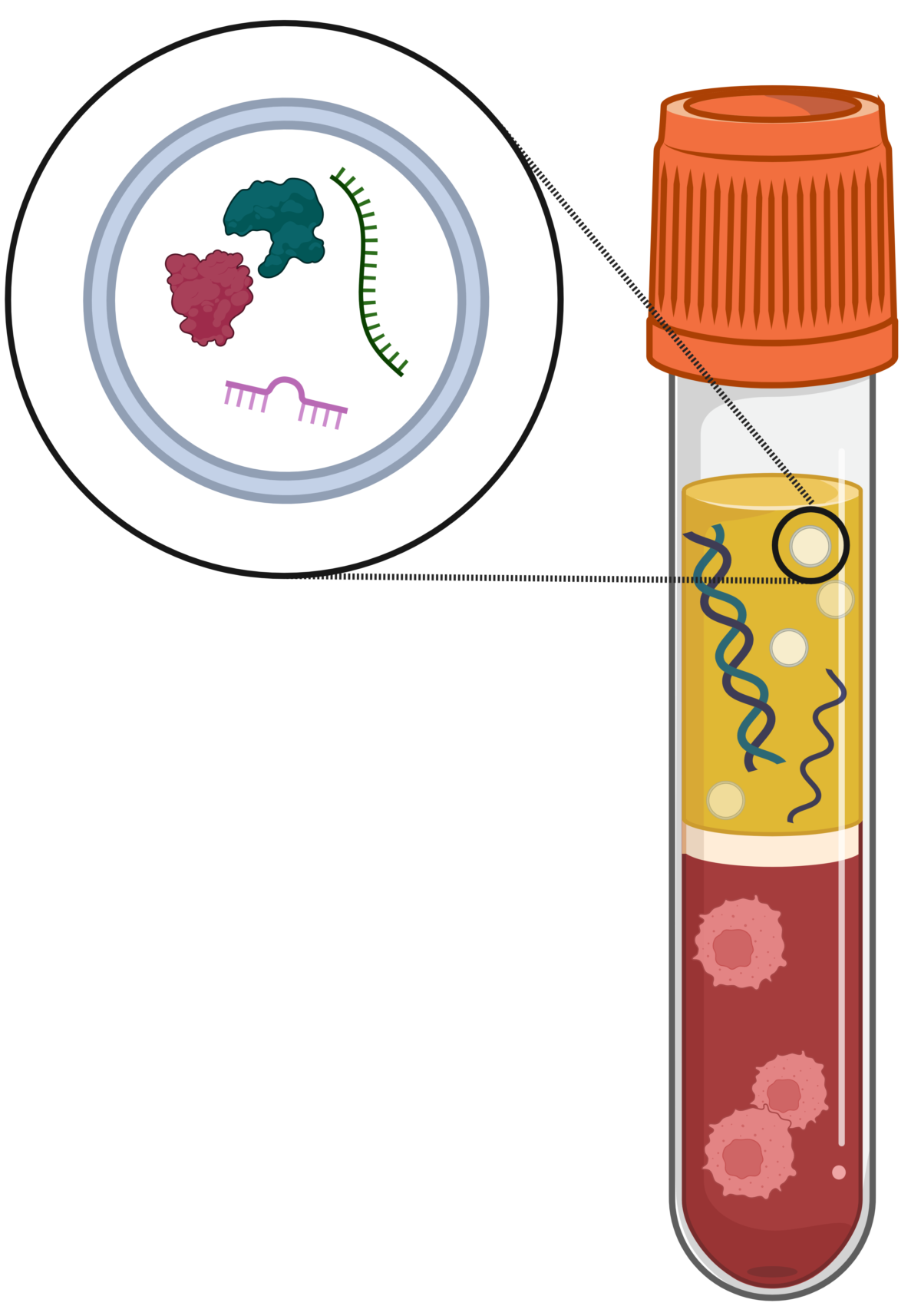
Novel biomarker panel for early detection of ovarian cancer
Engineering tumor cellular compartmentalization with tumor-on-a-chip
Tumors are compartmentalized encompassing a complex network of cellular and non-cellular components, including fibroblasts, imperfect vascularization, spatial heterogeneity in the distribution of nutrients, oxygen, and signaling molecules, and ECM remodeling, which collectively create a specialized niche supporting tumor growth. Unfortunately, there are key limitations with current preclinical models recapitulating these key cellular crosstalk and spatial organization. We have developed a multicompartmentalized microvascularized tumor-on-a-chip as a novel preclinical model that recapitulates those key components of the dynamic and compartmentalized tumors microenvironment to further evaluate their critical role in modulating drug resistance.
Plesselova S, Calar K, Axemaker H, Sahly E, Bhagia A, Faragher J, Fink D, de la Puente P. Multicompartmentalized microvascularized tumor-on-a-chip to study tumor-stroma interactions and drug resistance in ovarian cancer. Cel. Mol. Bioeng. (2024). https://doi.org/10.1007/s12195-024-00817-y
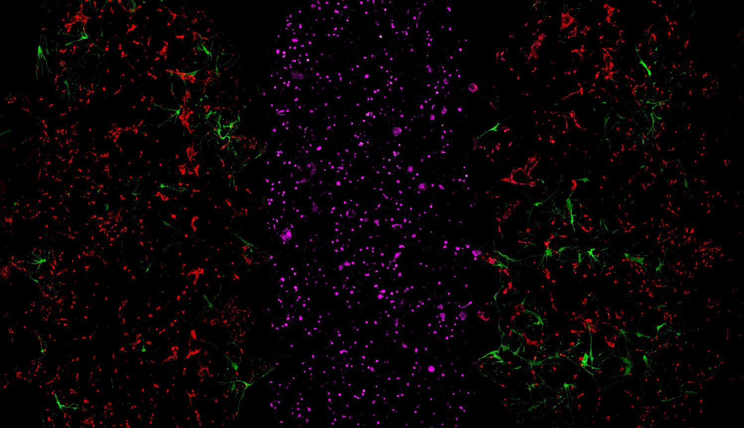
Tumor-on-a-chip with tri-culture design including cancer, cancer associated fibroblast and HUVECs
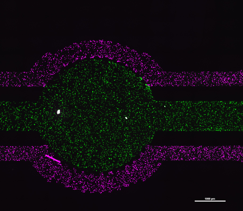
Tumor-on-a-chip with tumor compartmentalization
